Hindrance Coefficient of the Vitreous Humor
Background
The vitreous humor is a gel-like material consisting of 99% water, 0.9% salts and the remaining is a network of collagen and hyaluronic acid. This network characterizes the porous structure of the vitreous humor. In a normal eye, water percolates though the vitreous humor, and carries with it the dissolved substances. With the introduction of drugs into the vitreous, these drugs will experience convective
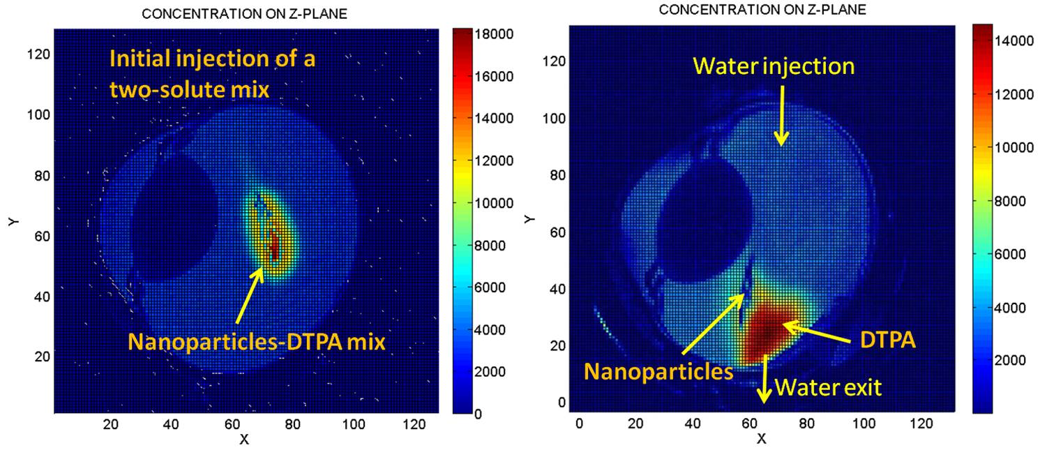
Fig. 1: ‘Before’ and ‘after’ MRI views of dual constrast-agent distribution before and after water flow in the vitreous humor of the ex vivo bovine eye. Water flow pushes the Gd-DTPA farther than the nanoparticles.
transport besides diffusion. However, with large molecules, the proper implementation of convective transport in gel portion of the vitreous requires the introduction of a resistive parameter (known as the Staverman filtration coefficient). We shall refer to it as the hindrance coefficient. This is necessary since without it the theory assumes the convective transport of the large molecule solute to be at the same rate as the solvent, aside from diffusion, and can overestimate the transport rate depending on the sizes of macromolecules. We have demonstrated in earlier studies that with very high water flow rates in ex vivo bovine eyes, while the water pushes the surrogate, significant residue of the latter remains. In particular, we introduced a mix of Gd-DTPA and gadolinium-based nanoparticles (Biopal), and injected the mix into the middle of the vitreous region of the eye (see image on the left in Figure 1). This was followed by an intense flow of water (much higher than the physiological flow for illustration purposes), injected by a syringe pump. At the diametrically opposite end of the eye, a drainage slit was cut. After several hours of pumping, the eye was imaged again (right image). It is seen that the nanoparticles lag behind the Gd-DTPA, clearly indicating the hindrance effect experienced by the nanoparticles.
Current Work
Preliminary work has been started with an extracted bovine vitreous by placing it in a clear cylindrical chamber (see Figure 2) and subjecting it to gravity-fed saline. The entire bovine vitreous was extracted and placed in the cylindrical chamber of the cell. At the start of the experiment, 50 micron colored microparticles were injected into the vitreous, and vitreous chamber was connected to slightly pressurized saline from an elevated source (60 cm) at one end, while the vitreal liquid was allowed to drain at the other. The microparticles in the shape of an elongated bolus were observed to migrate in the direction of the flow. An initial quantification indicated that the nanoparticles moved slower than the water flow. There was no observed diffusion of the microparticles, and the migration was entirely convective.
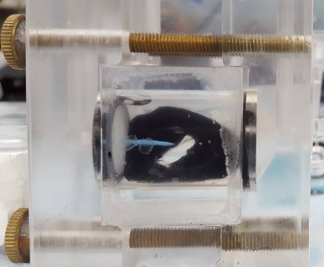
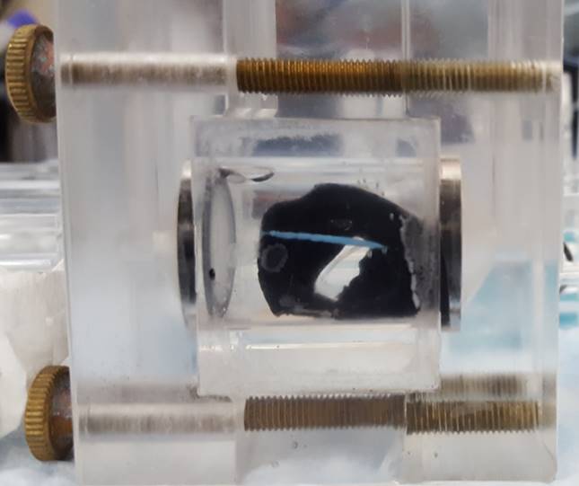
Figure 2: Microparticles in extracted bovine vitreous subjected to water flow (left to right). The vitreous with the hyaloid membrane intact is placed in the cylindrical chamber. Saline is fed on the left from an 80-cm elevated source, and allowed to exit at the right. After 89 minutes of water flow through the vitreous, the elongated nanoparticles bolus is seen to migrate in the direction of flow. There is almost no diffusion